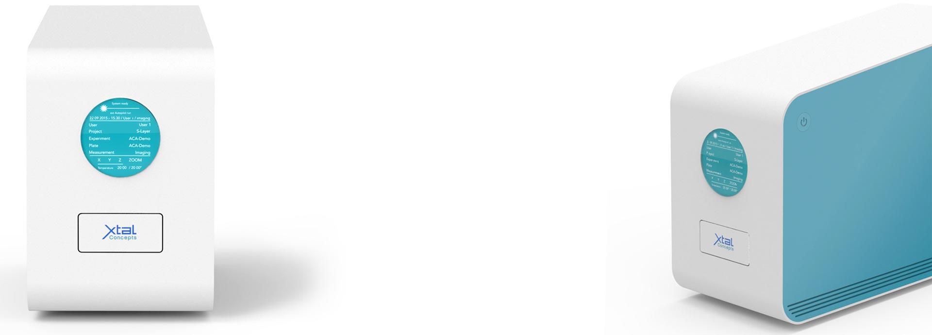
SpectroLight 600/610
is a powerful benchtop instrument capable of fully automated Dynamic Light Scattering (DLS) particle sizing in unsurpassed small sample volumes (0.08 – 1 µl). The sample carrier is a standard 96-well plate, available from many laboratory suppliers. These features make the SpectroLight 600 ideal for many laboratory applications such as sample quality or stability analysis, buffer screening and many more. In addition to the DLS, SpectroLight provides a fully integrated laboratory microscope for visual inspection of samples, which has proven to be a great help in detecting colloidal or solid contaminants. In addition, the imaging capabilities of the SpectroLight 600 extend its range of applications. It can be used as a fully automated imaging system for rapid crystal identification with optional UV imaging.
Make your sample fit for modern analysis techniques
Once a sample has been identified as monodisperse, it is already qualified for almost all subsequent structural determination methods, as a uniform population of proteins in solution is essential for highly reliable data.
One for All
SpectroLight 600 offers unrivalled versatility in its range of applications. It is not only the most sample efficient high throughput microplate based dynamic light scattering instrument on the market, but also a full featured imaging system. Optional configurations are available to meet specific customer requirements.
Buffer screening
In situ DLS is a rapid and reliable method for scoring protein responses to a multitude of buffer conditions based on the particle size.
Unmatched small Sample Volume
A key feature of sample efficiency is the ability to perform measurements on the smallest possible volumes. With standard volumes ranging from 80 to 500 nl/well in microtiter plates, SpectroLight 600 enables DLS measurements on unrivalled small sample volumes.
Non-Invasiveness
A non-invasive and direct detection method that registers moving particles in solution without changing them, that is close to the optimum detection priciple generally conceivable. And exactly this is what in situ DLS is about.
Particle Size Determination
Particle sizes can be determined in droplets, which is called "in situ" by applying a laser beam directly into a sample drop located in a well of a plate. Particles can be measured in a range of 1 nm up to a few micrometres. That closes the gap, between the vis-microscope observable and the molecular world. Based on hydrodynamics, particle size information can be obtained with +/- 2% accuracy and determined even when they coexist in a mixture of several populations.
Workflow Monitoring
Quick data acquisition and a direct output on small aliquots are enabled by situ DLS and these are key features of an "all over monitoring" approach. Keeping a sample monodisperse throughout all steps indicates a "feeling good" situation for the protein.
Applications (PDF files)
– Unmatched small Sample Volume
– Monomer/Oligomer Distinction
– Robustness of in situ DLS
– Sample Concentrating
– Cryo EM-Applications
REFERENCES
James Birch , Danny Axford , James Foadi , Arne Meyer , Annette Eckhardt , Yvonne Thielmann , Isabel Moraes
The fine art of integral membrane protein crystallisation, Methods 2018 Sep 1;147:150-162. doi: 10.1016/j.ymeth.2018.05.014. Epub 2018
Karsten Dierks, Arne Meyer, Howard Einspahr and Christian Betzel
Dynamic Light Scattering in Protein Crystallization Droplets: Adaptations for Analysis and Optimization of Crystallization Processes, Cryst. Growth Des., 2008, 8 (5), pp 1628–1634
Dominik Oberthuer, Emilio Melero-García, Karsten Dierks, Arne Meyer, Christian Betzel, Alfonso Garcia-Caballero and Jose A. Gavira
Monitoring and Scoring Counter-Diffusion Protein Crystallization Experiments in Capillaries by in situ Dynamic Light Scattering,
PLoS ONE 7(6): e33545. doi:10.1371/journal.pone.0033545
Arne Meyer, Karsten Dierks, Rana Hussein, Karl Brillet, Hevila Brognaro, and Christian Betzel
Systematic analysis of protein-detergent complexes applying dynamic light scattering to optimize solutions for crystallization trials,
Acta Crystallogr F Struct Biol Commun. 2015 Jan 1; 71(Pt 1): 75–81