Powered by in-plate DLS
What does it mean? Well, we made some essential contributions to two scientific publications in which in-plate DLS data were collected during device demonstrations in 2026. This resulted in an XtalConcepts member becoming one of the paper's co-authors.
How is something like that possible? The answer lies in the unique combination of features of our non-invasive in-plate dynamic light scattering (DLS) systems. This opens up a whole new level of analytical potential. It enables applications that were previously impossible.
The key features that made this contibutions possible are:
- 96 wells enable DLS scans to be run on multiple samples over long time periods.
- the exceptionally small sample volume, together with the non-invasive
- Accuracy of this method that allows for experiments to provide answers to long-standing questions
- Long-term measurements on the same sample
The two examples from 2026 are highlighted:
The first is the development of targeted protein degradation by proteolysis-targeting chimeras (PROTACs) recruiter assay against the main SARS-CoV-2 protease, Mpro (manuscript submitted [1]):
Identification of the PROTAC (variat BT153) mediated ternary complex of the DDB1/CRBN ubiquitination complex with the SARS-CoV-2 Protease Mpro by a newly developed Recruiter Assay-Precursor:
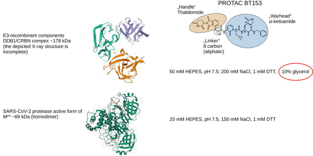
Below: In-plate DLS-PROTAC recruiter assay precursor: Loading scheme and evaluation. Buffer conditions were loaded in columns. Some formulations and other tests were loaded row-wise.
Row A is particularly important since the DDB1/CRBN and Mpro formulations were not identical: the DDB1/CRBN formulation contains 10% glycerol, resulting in significantly different viscosity.
Since absolute sizes were used to identify the ternary complex, viscosity had to be determined by applying 100 nm LATEX-nanospheres and using a SpectroLight 600 microviscosimeter.
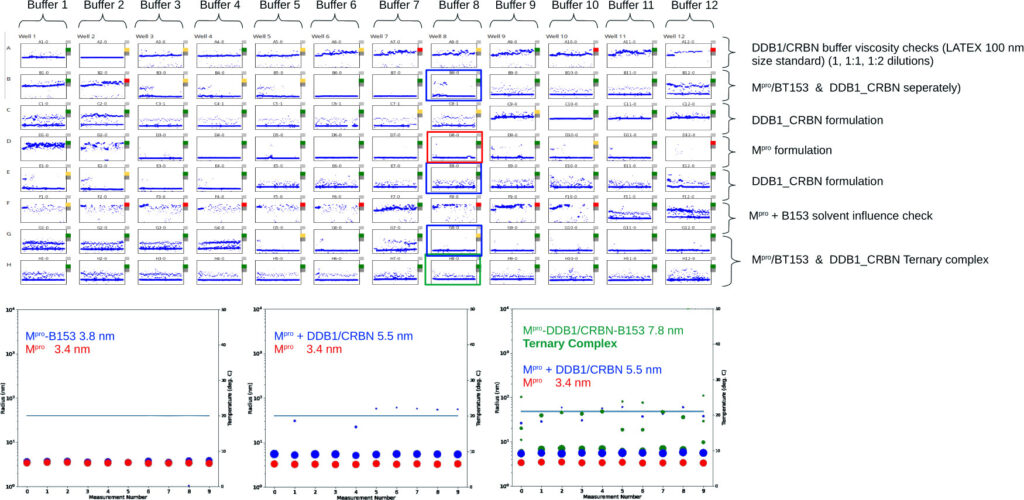
The BT153-mediated ternary complex (Rh 7.8 nm) exceeds the larger DDB1/CRBN component (Rh 5.5 nm). This is a typical outcome of successful non-covalent cross-linking with a 1:1:1 subunit and PROTAC stoichiometry.
Another example of co-authorship in 2026, following a demonstration of a device, involved replacing an incompatible detergent used in an activity assay with a new one for the lysosomal enzyme complex of the signal peptide peptidase (SPPL).
Identifying a suitable detergent is exceptionally challenging since the active site is located within the transmembrane region. Solubilisation by detergents often results in the rearrangement of the multi-helical transmembrane region, cancelling out the enzymatic activity of this class of enzymes. This challenge was overcome by maintaining the complex's absolute size at a sub-nanometre level, thus avoiding helix-randomising detergents.
Examples of the detergent screen are shown below: Identification was based on a comparison between the original, incompatible detergent and the new one.
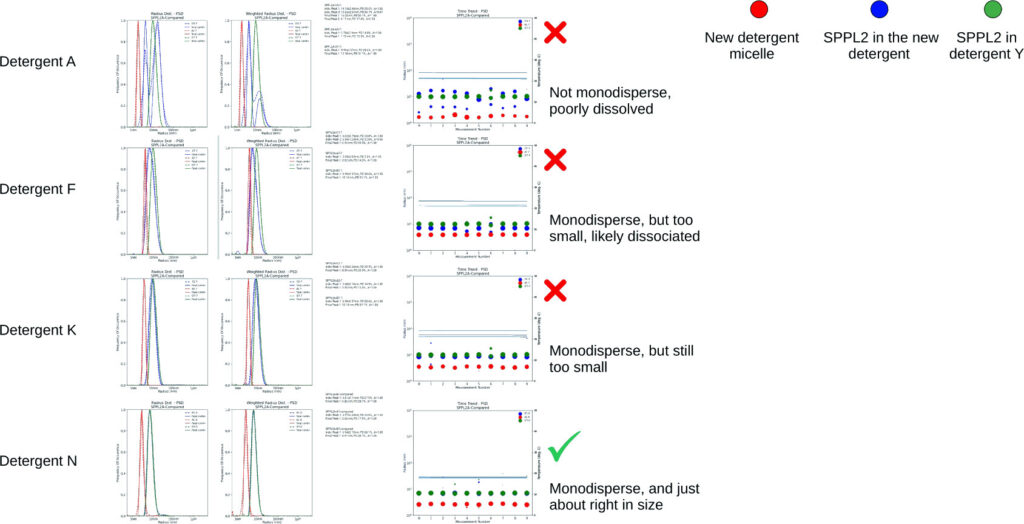
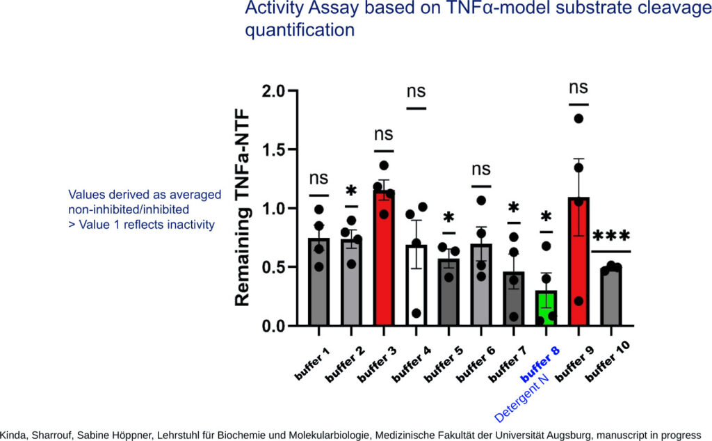
Detergent N was in almost exact superposition in an overlay of the size plots. The applied activity assay was recently introduced [2], and SPPL shows the strongest activity in detergent N. A second in-plate DLS run determined that the active form was tetrameric rather than monomeric [3].
Results of the SPPL activity assay:
These two examples from 2026 alone show the enormous impact that this technologie has, once it is implemented in the scientific workflow.
Please visit our website: XtalConcepts, in order to learn more about our powerful products.
Your XtalConcepts Team
References:
1. Y-T. Chen, K. Zhang, A. Meyer, M. Brönstrup et al. An antiviral PROTAC against the main protease of SARS-CoV-2 acts through monoubiquitin-mediated lysosomal and proteasomal degradation 2026 (submitted 2026 to Cell Chemical Biology), in cooperation with Prof. Dr. Mark Brönstrup Chemische Biologie (CBIO), HZI - Helmholtz-Zentrum für Infektionsforschung GmbH, Inhoffenstraße 7, 38124 Braunschweig
2. Sharrouf et al. (2024)In vitro cleavage of tumor necrosis factor α (TNFα) by Signal-Peptide-Peptidase-like 2b (SPPL2b) resembles mechanistic principles observed in the cellular context, Chemico-Biol. Interactions, Volume 395, 25 May 2024, 111006
3. K. Sharrouf, A. Meyer, S. Höppner et al. (2026, Manuscript in preparation)
In cooperation with Prof. Sabine Höppner, Faculty of Medicine, University of Augsburg, Universitätsstr. 2, 86159 Augsburg